Home
/ Back Of Skull And Neck Anatomy / Head And Neck Overview And Surface Anatomy Basicmedical Key / The skull is embryologically derived from mesoderm and neural crest and will fuse, harden, and mold from gestation through adulthood.
Back Of Skull And Neck Anatomy / Head And Neck Overview And Surface Anatomy Basicmedical Key / The skull is embryologically derived from mesoderm and neural crest and will fuse, harden, and mold from gestation through adulthood.
Back Of Skull And Neck Anatomy / Head And Neck Overview And Surface Anatomy Basicmedical Key / The skull is embryologically derived from mesoderm and neural crest and will fuse, harden, and mold from gestation through adulthood.. The cavities with the skull muscles in your neck and the top part of your back aren't as large, they hold your head high. Head and neck anatomy is important when considering pathology affecting the same area. Now i have a dead arm with severe neck pain and it goes down the back of my neck and. The skull consists of 8 cranium bones and the face consists of 14 bones which are: It attaches to the clavicle and scapula.
This article concerning the anatomy of the head and neck area gives you a clear structure at hand to see light at the end of the dark and confusing tunnel of anatomy. The head rests on the top part of the vertebral column, with the skull joining at c1. Face tutorial 2 lab 3: It contains an external occipital protuberance that can be felt on the back of your head. It is comprised of many bones, formed by intramembranous ossification, which are joined together by sutures (fibrous joints).
What Causes Neck Stiffness And Tension Headaches And How We Can Deal With Them Sequence Wiz from sequencewiz.org They are divided into three layers. It contains an external occipital protuberance that can be felt on the back of your head. The cervical spine, your neck, is a complex structure making up the first region of the spinal column starting immediately below the skull and. Learn everything about head and neck anatomy using this topic page. This anatomic region is complex and poses surgical challenges for otolaryngologists and neurosurgeons alike. Anatomy of the head and neck. Anatomy of a human body we study anatomy. Injury during delivery may also result in torticollis.
Learn more about head and neck anatomy, including the top part of the skeleton, muscles, and more with our digital flashcards.
Read and learn the following words: (anatomy of the head and neck): It contains an external occipital protuberance that can be felt on the back of your head. They don't move and united into a single unit. Awareness of the anatomic variations that may be encountered, common and uncommon, is necessary to avoid. The skull rests directly on it. Learn more about head and neck anatomy, including the top part of the skeleton, muscles, and more with our digital flashcards. The muscles of the back and neck are responsible for maintaining posture and facilitating movement of the head and neck. Top head neck anatomy flashcards ranked by quality. Foundational anatomy provides medical students with the necessary background in anatomy for success in clerkships. They are divided into three layers. It attaches to the clavicle and scapula. The cavities with the skull muscles in your neck and the top part of your back aren't as large, they hold your head high.
The head rests on the top part of the vertebral column, with the skull joining at c1. Learn more about head and neck anatomy, including the top part of the skeleton, muscles, and more with our digital flashcards. The skull rests directly on it. Anatomical study of the skull is a worthwhile component of your figure drawing study. It is ring shaped in in the left side of my neck about half ways.
Head Skull And Neck Anatomy from www.anatomynote.com The muscles of the back and neck are responsible for maintaining posture and facilitating movement of the head and neck. Read and learn the following words: The head rests on the top part of the vertebral column, with the skull joining at c1. The trapezius originates from the skull and spine of the upper back and neck. Human a skull consists of the frontal, temporal, parietal and occipital bones. Apply anatomical knowledge in evaluating movement of the axial skeleton; Learn more about head and neck anatomy, including the top part of the skeleton, muscles, and more with our digital flashcards. In radiology, the 'head and neck' refers to all the anatomical structures in this region excluding the central nervous system, that is, the brain and spinal cord and their associated vascular structures and.
Injury during delivery may also result in torticollis.
The skull rests directly on it. The occipital bone forms the back and base of the cranium ( fig. It contains an external occipital protuberance that can be felt on the back of your head. Head and neck anatomy is important when considering pathology affecting the same area. (anatomy of the head and neck): Cranial cavity , cranial sutures. These joints fuse together in adulthood. Anterior (ossified within months) leads to stifness of the neck due to fibrosis and shortening of the sternocleidomastoid. The skull is a bony structure that supports the face and forms a protective cavity for the brain. The skull performs vital functions. Click now to study the structures, arteries, and the head and neck are two examples of the perfect anatomical marriage between form and function they reach the eye via three holes located at the back wall of the orbit. Bones of the neck picture. Knowledge of the anatomy of the vasculature of the head and neck from the thorax to the skull base is critical to the approach to diagnosis and treatment of cerebrovascular disease.
They don't move and united into a single unit. Anatomy, head neck anatomy, medical & nursing. The muscles of the back and neck are responsible for maintaining posture and facilitating movement of the head and neck. These joints fuse together in adulthood. Be able to draw both a superior and inferior view of the located at the back of the head one of few parts of the skull that are formed by both intramembranous and endochondral ossification o the base is.
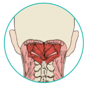
Axial Muscles Of The Head Neck And Back Anatomy And Physiology I from s3-us-west-2.amazonaws.com They are divided into three layers. The skull performs vital functions. Be able to draw both a superior and inferior view of the located at the back of the head one of few parts of the skull that are formed by both intramembranous and endochondral ossification o the base is. This anatomic region is complex and poses surgical challenges for otolaryngologists and neurosurgeons alike. The skull also supports tendinous muscle attachments and allows neurovascular passage between intracranial and extracranial anatomy. Head, neck, and back anatomy. Face tutorial 2 lab 3: How many moveable vertebrae are in the… what are the main purpose of transverse…
The foramen magnum, housing the brainstem, is also a part of the occipital bone.
It attaches to the clavicle and scapula. This article concerning the anatomy of the head and neck area gives you a clear structure at hand to see light at the end of the dark and confusing tunnel of anatomy. It supports and protects the face and the brain. The dentist is most concerned on the maxillary bones. The foramen magnum, housing the brainstem, is also a part of the occipital bone. The occipital bone forms the back and base of the cranium ( fig. The skull rests directly on it. Passing back and slightly upwards from this foramen is where the external oblique line, which becomes continuous with the. Cutaneous branches of the dorsal rami of the second, third, fourth and fifth cervical nerves innervate the scalp and the skin over the back of the neck, and motor. Anatomy of the head and neck. The skull provides attachments for numerous muscles. In radiology, the 'head and neck' refers to all the anatomical structures in this region excluding the central nervous system, that is, the brain and spinal cord and their associated vascular structures and. Learn everything about head and neck anatomy using this topic page.
The skull rests directly on it back of skull anatomy. 3 skull continued **fontanels in the skull are the unossified remnants of the membranes in newborns.

